• Sale!

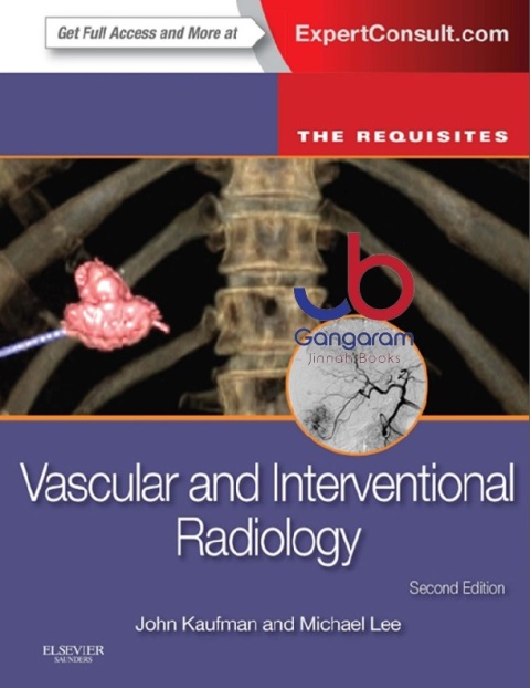
    Vascular and Interventional Radiology: The Requisites (The Core Requisites) 2nd Edition

    by John A. Kaufman MD MS FSIR FAHA FCIRSE EBIR (Author), Michael J. Lee MSc FRCPI FRCR FFR(RCSI) FSIR EBIR (Author)

    Get the essential tools you need to make an accurate diagnosis with Vascular and Interventional Radiology: The Requisites! This bestselling volume delivers the conceptual, factual, and interpretive information you need for effective clinical practice in vascular and interventional radiology, as well certification and recertification review. Master core knowledge the easy and affordable way with clear, concise text enhanced by at-a-glance illustrations, boxes, and tables ? all completely rewritten to bring you up to date with today?s state of the art in vascular and interventional radiology.

    "... a volume that should retain its utility for several years to come, both as a primer for radiology trainees and fellows at the start of their IR training and as a reference for more experienced interventionalists." Reviewed by Dr Simon Padley and Dr Narayanan Thulasidasan on behalf of RAD Magazine, April 2015

    Understand the basics with a comprehensive yet manageable review of the principles and practice of vascular and interventional radiology. Whether you’re a resident preparing for exams or a practitioner needing a quick-consult source of information, Vascular and Interventional Radiology is your guide to the field. Master the latest techniques for liver-directed cancer interventions; arterial and venous interventions including stroke therapy; thoracic duct embolization; peripheral arterial interventions; venous interventions for thrombosis and reflux; percutaneous ablation procedures; and much more. Prepare for the written board exam and for clinical practice with critical information on interventional techniques and procedures. Clearly visualize the findings you’re likely to see in practice and on exams with vibrant full-color images and new vascular chapter images. Access the complete, fully searchable text and downloadable images online with Expert Consult.

    You can buy this product at Gangaram Jinnah Medical Books Shop for home delivery and Cash on delivery to all over Pakistan. All kind of medical books are available.
  • Sale!

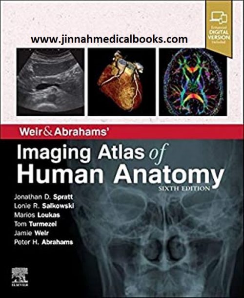
    Weir & Abrahams’ Imaging Atlas of Human Anatomy 5th Edition

    Original price was: 800 ₨.Current price is: 700 ₨.
    Weir & Abrahams' Imaging Atlas of Human Anatomy 5th Edition by Jonathan D. Spratt MA (Cantab) FRCS (Eng) FRCR (Author), Lonie R Salkowski MD (Author), Marios Loukas MD PhD (Author), Tom Turmezei BMBCh MA MPhil FRCR (Author), Jamie Weir MB BS FRCP(Ed) FRCR (Author) Imaging is ever more integral to anatomy education and throughout modern medicine. Building on the success of previous editions, this fully revised fifth edition provides a superb foundation for understanding applied human anatomy, offering a complete view of the structures and relationships within the body using the very latest imaging techniques. It is ideally suited to the needs of medical students, as well as radiologists, radiographers and surgeons in training. It will also prove invaluable to the range of other students and professionals who require a clear, accurate, view of anatomy in current practice.
    • Fully revised legends and labels and over 80% new images – featuring the latest imaging techniques and modalities as seen in clinical practice
    • Covers the full variety of relevant modern imaging – including cross-sectional views in CT and MRI, angiography, ultrasound, fetal anatomy, plain film anatomy, nuclear medicine imaging and more – with better resolution to ensure the clearest anatomical views
    • Unique new summaries of the most common, clinically important anatomical variants for each body region – reflects the fact that around 20% of human bodies have at least one clinically significant variant
    • New orientation drawings – to help you understand the different views and the 3D anatomy of 2D images, as well as the conventions between cross-sectional modalities
    • Now a more compete learning package than ever before, with superb new BONUS electronic enhancements embedded within the accompanying eBook, including:
      • Labelled image ‘stacks’ - that allow you to review cross-sectional imaging as if using an imaging workstation
      • Labelled image ‘slide-lines’ - showing features in a full range of body radiographs to enhance understanding of anatomy in this essential modality
      • Self-test image ‘slideshows’ with multi-tier labelling - to aid learning and cater for beginner to more advanced experience levels
      • Labelled ultrasound videos - bring images to life, reflecting this increasingly clinically practiced technique
      • Questions and answers accompany each chapter - to test your understanding and aid exam preparation
      • 34 pathology tutorials – based around nine key concepts and illustrated with hundreds of additional pathology images, to further develop your memory of anatomical structures and lead you through the essential relationships between normal and abnormal anatomy
  • Sale!

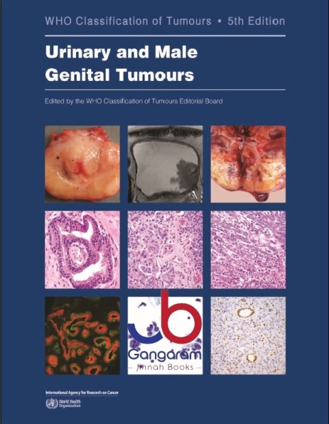
    WHO classification of tumours of the urinary system and male genital organs: 8 (World Health Organization classification of tumours, 9)

    by International Agency for Research on Cancer (Author), H. Moch (Editor)

    Urinary and Male Genital Tumours is Volume 8 in the 5th edition of the WHO series on the classification of human tumors.

    This series (also known as the WHO Blue Books) is regarded as the gold standard for the diagnosis of tumors and comprises a unique synthesis of histopathological diagnosis withdigital and molecular pathology. These authoritative and concise reference books provide indispensable international standards for anyone involved in the care of patients with cancer or in cancer research, underpinning individual patient treatment as well as research into all aspects of cancer causation, prevention, therapy, and education.

    What's new in this edition?

    The 5th edition, guided by the WHO Classification of Tumours Editorial Board, will establish a single coherent cancer classification presented across a collection of individual volumes organized on the basis of anatomical site (digestive system, breast, soft tissue and bone, etc.) and structured in a systematic manner, with each tumor type listed within a taxonomic classification: site, category, family (class), type, and subtype. In each volume, the entities are now listed from benign to malignant and are described under an updated set of headings, including histopathology, diagnostic molecular pathology, staging, and easy-to-read essential and desirable diagnostic criteria.

    Who should read this book?

    Pathologists Urologists Radiologists Medical oncologists Radiation oncologists Surgeons Oncology nurses Cancer researchers Epidemiologists Cancer registrars This volume: Prepared by 181 authors and editors Contributors from around the world More than 900 high-quality images More than 3600 references

    You can buy this product at Gangaram Jinnah Medical Books Shop for home delivery and Cash on delivery to all over Pakistan. All kind of medical books are available.
Sale