• Sale!

    Textbook of Clinical Neuroanatomy Vishram SIngh 3rd Edition

    Original price was: 800 ₨.Current price is: 650 ₨.

    Textbook of Clinical Neuroanatomy Vishram Singh 3rd Revised 

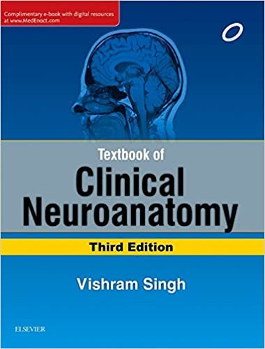
    published by Elsevier, is primarily compiled for undergraduate medical and dental students. It is an authoritative reference source for postgraduates and practicing neurologists and neurosurgeons. The salient features of the book include the following: all chapters are revised and updated, including details on cranial nerves and their lesions, blood supply and cerebrovascular accidents, motor and sensory disorders; new line diagrams, and real life photographs and MRI scans; simple, to-the-point, easy-to-understand exam-oriented text; numerous, four-coloured, large sized, and easy-to-draw diagrams; and the text offers unique problem-based clinical and functional perspective.
    • ISBN-10: 8131244644
    • ISBN-13: 978-8131244647
     
  • Sale!
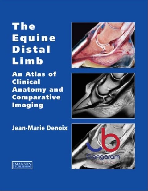
    The Equine Distal Limb: An Atlas of Clinical Anatomy and Comparative Imaging 1st Edition by Jean-Marie Denoix (Author) Jean-Marie Denoix is the world’s leading equine musculoskeletal system anatomist and has become one of the foremost equine diagnostic ultrasonographers. There is therefore nobody better to compile a reference atlas of the clinical anatomy of the foot, pastern and fetlock, correlated with images obtained by radiography, diagnostic ultrasonography and magnetic resonance imaging. Advanced imaging techniques require in depth knowledge of anatomy for accurate interpretation and especially when using magnetic resonance imaging this must be a 3-dimensional concept of anatomy. This book provides a unique contribution and is an invaluable reference text. The image quality is extraordinarily high and the multiple views of each area of the distal limb provide an extremely detailed evaluation. Each part opens with a characteristically superb anatomical drawing by the author. Each spread of pages deals with a single dissection viewed by means of colour photographs, labelled black and white equivalents, plus x-rays, ultrasound and MRI scans as required. The book is designed for maximum clarity using a generous page size. The atlas is essential for anybody involved in detailed anatomical study, complex lameness evaluation or advanced imaging techniques. You can buy this product at Gangaram Jinnah Medical Books Shop for home delivery and Cash on delivery to all over Pakistan. All kind of medical books are available.  
  • Sale!
    by Matthew A. Mulvey (Author), David J. Klumpp (Author), Ann E. Stapleton (Author) A comprehensive overview of clinically important infections of the urinary tract Urinary tract infections (UTIs) continue to rank among the most common infectious diseases of humans, despite remarkable progress in the ability to detect and treat them. Recurrent UTIs are a continuing problem and represent a clear threat as antibiotic-resistant organisms and infection-prone populations grow. Urinary Tract Infections: Molecular Pathogenesis and Clinical Management brings the scientific community up to date on the research related to these infections that has occurred in the nearly two decades since the first edition. The editors have assembled a team of leading experts to cover critical topics in these main areas: clinical aspects of urinary tract infections, including anatomy, diagnosis, and management, featuring chapters on the vaginal microbiome as well as asymptomatic bacteriuria, prostatitis, and urosepsis the origins and virulence mechanisms of the bacteria responsible for most UTIs, including uropathogenic Escherichia coli, Proteus mirabilis, and Klebsiella pneumoniae the host immune response to UTIs, the rise of antibiotic-resistant strains, and the future of therapeutics This essential reference serves as both a resource and a stimulus for future research endeavors for anyone with an interest in understanding these important infections, from the classroom to the laboratory and the clinic. You can buy this product at Gangaram Jinnah Medical Books Shop for home delivery and Cash on delivery to all over Pakistan. All kind of medical books are available.
  • Sale!
    Author: Hoballah, Jamal J. This must-have manual for surgical residents and vascular surgery trainees is copiously illustrated with more than 250 original line drawings. The author presents in a step- by-step fashion the techniques for all common vascular reconstructions, basics of exposure and access, as well as invaluable technical pearls and tips. The text provides the surgical trainee with the opportunity to review and practice the necessary vascular reconstructions before entering the operating room. The illustrations and clear instructions make this a manual that is referred to again and again throughout the surgical residency. You can buy this product at Gangaram Jinnah Medical Books Shop for home delivery and Cash on delivery to all over Pakistan. All kind of medical books are available.  
  • Sale!
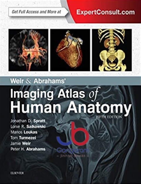
    by Jonathan D. Spratt MA (Cantab) FRCS (Eng) FRCR (Author), Lonie R Salkowski MD (Author), Marios Loukas MD PhD (Author), Tom Turmezei BMBCh MA MPhil FRCR (Author), Jamie Weir MB BS FRCP(Ed) FRCR (Author), Peter H. Abrahams MBBS FRCS(ED) FRCR DO(Hon) FHEA (Author) Imaging is ever more integral to anatomy education and throughout modern medicine. Building on the success of previous editions, this fully revised fifth edition provides a superb foundation for understanding applied human anatomy, offering a complete view of the structures and relationships within the body using the very latest imaging techniques. It is ideally suited to the needs of medical students, as well as radiologists, radiographers and surgeons in training. It will also prove invaluable to the range of other students and professionals who require a clear, accurate, view of anatomy in current practice. Fully revised legends and labels and over 80% new images – featuring the latest imaging techniques and modalities as seen in clinical practice Covers the full variety of relevant modern imaging – including cross-sectional views in CT and MRI, angiography, ultrasound, fetal anatomy, plain film anatomy, nuclear medicine imaging and more – with better resolution to ensure the clearest anatomical views Unique new summaries of the most common, clinically important anatomical variants for each body region – reflects the fact that around 20% of human bodies have at least one clinically significant variant New orientation drawings – to help you understand the different views and the 3D anatomy of 2D images, as well as the conventions between cross-sectional modalities Now a more compete learning package than ever before, with superb new BONUS electronic enhancements embedded within the accompanying eBook, including: Labelled image ‘stacks’ - that allow you to review cross-sectional imaging as if using an imaging workstation Labelled image ‘slide-lines’ - showing features in a full range of body radiographs to enhance understanding of anatomy in this essential modality Self-test image ‘slideshows’ with multi-tier labelling - to aid learning and cater for beginner to more advanced experience levels Labelled ultrasound videos - bring images to life, reflecting this increasingly clinically practiced technique Questions and answers accompany each chapter - to test your understanding and aid exam preparation 34 pathology tutorials – based around nine key concepts and illustrated with hundreds of additional pathology images, to further develop your memory of anatomical structures and lead you through the essential relationships between normal and abnormal anatomy You can buy this product at Gangaram Jinnah Medical Books Shop for home delivery and Cash on delivery to all over Pakistan. All kind of medical books are available.  
  • Sale!

    Wheelers Dental Anatomy, Physiology And Occlusion

    Original price was: 2,895 ₨.Current price is: 2,500 ₨.
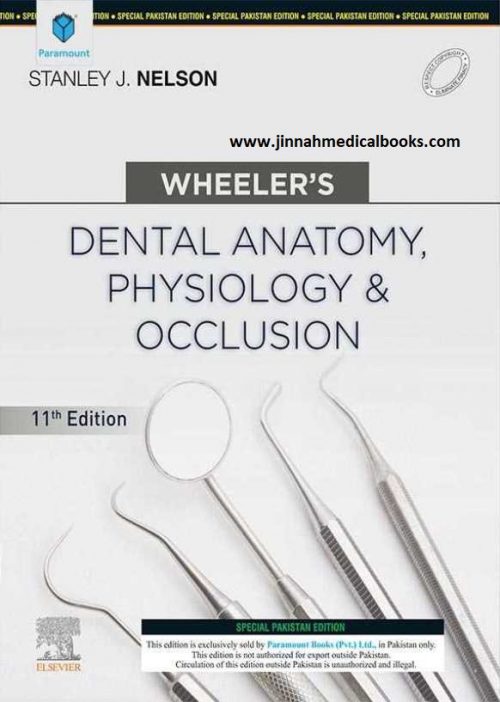
    Wheeler's Dental Anatomy, Physiology and Occlusion by Stanley J. Nelson   Successfully learn to apply dental anatomy to the practice of dentistry with Wheeler’s Dental Anatomy, Physiology, and Occlusion. Updated and visually enhanced this market-leading dental text expands its focus on clinical applications. Dozens of online 360-degree and 3-D tooth animations to give you an unparalleled view of dentitions, pulp formation, the sequence of eruptions, and countless clinical considerations. Every text purchase also comes with access to the companion Expert Consult website ― where you can reference the entire text electronically, as well as engage with a wealth of learning activities, animations, and hands-on resources to better understand and retain the information covered in the text. In all, this proven learning package offers all the up-to-date information, best practices, and tools to prepare you for the dental anatomy and occlusion section of the exams and ensure long-term clinical success.
    • More than 800 full-color images include detailed, well-labeled anatomical illustrations as well as clinical photographs.
    • Bolded key terms draw students’ attention to essential terminology.
    • Practical appendices include Review of Tooth Morphology with a concise review of tooth development from in utero to adolescence to adulthood; and Tooth Traits of the Permanent Dentition with tables for each tooth providing detailed information such as tooth notation, dimensions, position of proximal contacts, heights, and curvatures.
    • 360-degree virtual reality and 3-D animations on Expert Consult help students refine their skills in tooth identification and examination.
    • Step-by-step videos on Expert Consult demonstrate occlusal adjustments.
    • Labeling exercises challenge students to identify tooth structures and facial anatomy with drag-and-drop labels.
    • Chapter on clinical applications includes practical applications and case studies to prepare students for exams. Topics include root planing and scaling; extraction techniques and forces; relationship of fillings to pulp form and enamel form; and occlusal adjustment of premature occlusal contacts and arch form in relationship to bite splint designs.
    • NEW! Learning objectives and pre-test questions at the start of every chapter focus students’ attention on the knowledge and critical thinking expectations for each chapter.
    • NEW! Full-color images have replaced many of the black and white images to give students a more vivid picture of clinical situations and procedures.
    • NEW! Updated information incorporates new research and visuals to ensure students are equipped with the latest best practices.
    • NEW! Access to the companion Expert Consult website includes hands-on exercises, animations, videos, quizzes, exams, and more to round out students’ learning experience and help ensure their ability to apply book content.
  • Sale!

    Wheelers Dental Anatomy, Physiology and Occlusion – Off Set

    Original price was: 500 ₨.Current price is: 300 ₨.
    Wheeler's Dental Anatomy, Physiology and Occlusion - Off Set by Stanley J. Nelson DDS MS   Successfully learn to apply dental anatomy to the practice of dentistry with Wheeler’s Dental Anatomy, Physiology, and Occlusion. Updated and visually enhanced this market-leading dental text expands its focus on clinical applications. Dozens of online 360-degree and 3-D tooth animations to give you an unparalleled view of dentitions, pulp formation, the sequence of eruptions, and countless clinical considerations. Every text purchase also comes with access to the companion Expert Consult website ― where you can reference the entire text electronically, as well as engage with a wealth of learning activities, animations, and hands-on resources to better understand and retain the information covered in the text. In all, this proven learning package offers all the up-to-date information, best practices, and tools to prepare you for the dental anatomy and occlusion section of the exams and ensure long-term clinical success.
    • More than 800 full-color images include detailed, well-labeled anatomical illustrations as well as clinical photographs.
    • Bolded key terms draw students’ attention to essential terminology.
    • Practical appendices include Review of Tooth Morphology with a concise review of tooth development from in utero to adolescence to adulthood; and Tooth Traits of the Permanent Dentition with tables for each tooth providing detailed information such as tooth notation, dimensions, position of proximal contacts, heights, and curvatures.
    • 360-degree virtual reality and 3-D animations on Expert Consult help students refine their skills in tooth identification and examination.
    • Step-by-step videos on Expert Consult demonstrate occlusal adjustments.
    • Labeling exercises challenge students to identify tooth structures and facial anatomy with drag-and-drop labels.
    • Chapter on clinical applications includes practical applications and case studies to prepare students for exams. Topics include root planing and scaling; extraction techniques and forces; relationship of fillings to pulp form and enamel form; and occlusal adjustment of premature occlusal contacts and arch form in relationship to bite splint designs.
    • NEW! Learning objectives and pre-test questions at the start of every chapter focus students’ attention on the knowledge and critical thinking expectations for each chapter.
    • NEW! Full-color images have replaced many of the black and white images to give students a more vivid picture of clinical situations and procedures.
    • NEW! Updated information incorporates new research and visuals to ensure students are equipped with the latest best practices.
    • NEW! Access to the companion Expert Consult website includes hands-on exercises, animations, videos, quizzes, exams, and more to round out students’ learning experience and help ensure their ability to apply book content.

Sale